
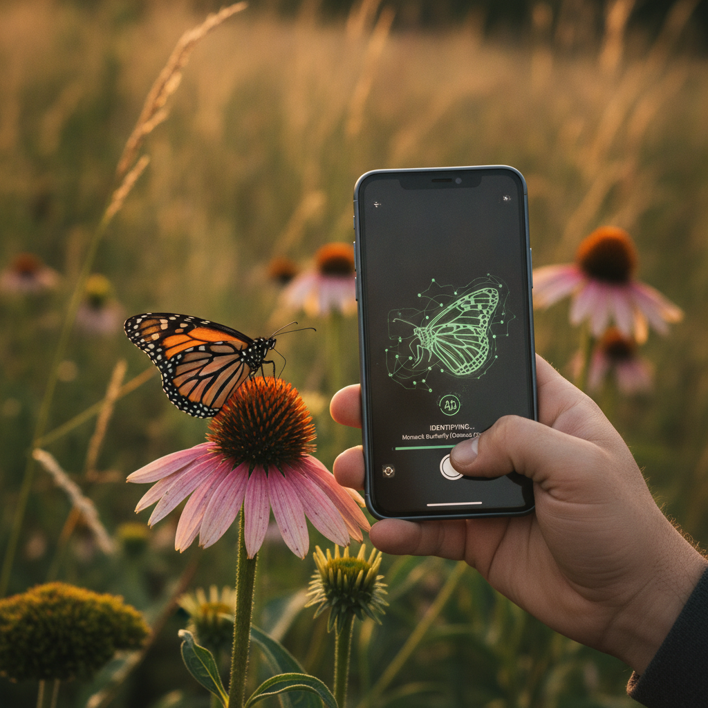
The Science Behind Visual Insect Identification
Every insect species represents millions of years of evolutionary refinement expressed through morphology — the physical form we photograph and analyze. Bug ID by photo is not guesswork dressed in technology. It is the translation of morphological characters that taxonomists have used since Linnaeus into pixels that algorithms and human eyes can evaluate. When I teach insect science to undergraduates, I emphasize that identification is a deductive process: each observed character narrows the possibility space until one species remains, or until you reach the resolution limit of your evidence.
Photographs capture a subset of morphological characters at a moment in time. That subset is often sufficient for family-level or genus-level identification and frequently adequate for species-level confirmation in groups with distinctive markings. It is rarely sufficient for cryptic species complexes that require genital dissection or molecular analysis. Understanding this resolution limit keeps bug ID by photo honest — powerful for education, ecology, and everyday curiosity, but not infallible for every taxonomic question.
The body plan of insects follows a modular architecture: head, thorax, and abdomen, with six legs and zero to two pairs of wings attached to the thorax. Variations on this theme produce the staggering diversity we observe. A photograph that clearly shows these modules and their ornamentation gives any identification system — human or artificial — the raw material for classification. A photograph that shows a blurry smear gives nothing useful regardless of software sophistication.
This guide walks through the morphological framework I use in teaching and field research, adapted for anyone performing bug ID by photo with a smartphone. You will learn which body regions matter most, how to recognize immature stages, how wing patterns and coloration function as taxonomic characters, and how to avoid the misidentifications that plague casual observers.
Key Body Regions for Photo-Based Bug ID
Taxonomists work through insects systematically, and your photographs should capture the same priority regions in order of diagnostic value.
The head carries mouthpart type, antennae structure, and eye configuration. Chewing mouthparts visible in mandibulate insects differ fundamentally from sucking mouthparts in true bugs and piercing-sucking mouthparts in butterflies and moths observed via coiled proboscis. Antennae segment count and shape — clubbed, serrated, thread-like, geniculate — separate families within orders. Compound eye size and spacing distinguish some fly and wasp groups. A clear head photograph, preferably oblique to show both antennae and mouthparts, resolves more identification questions than any other single view.
The thorax bears wings and legs and often displays critical coloration. The pronotum — the dorsal plate of the first thoracic segment — is prominently visible in beetles and true bugs and frequently bears species-specific markings. Scutellum shape and color separate true bug families. Wing attachment and venation patterns, visible when wings are spread or at rest, provide deep taxonomic information for Lepidoptera, Odonata, and Neuroptera. Leg spurs, tibial spines, and tarsal formulas matter for some groups, though phone cameras rarely capture tarsal detail without macro lenses.
The abdomen reveals segmentation, terminal appendages, and patterning. Cerci — paired appendages at the abdomen tip — identify many orthopterans and some beetles. Abdominal banding distinguishes wasp and bee species when thoracic characters are ambiguous. In larvae and caterpillars, abdominal prolegs and their arrangement on caterpillars separate butterfly from moth caterpillars with high reliability.
When performing bug ID by photo, ensure at least one image documents each major body region. Apps weight different characters differently, but comprehensive coverage maximizes accuracy across all algorithms.
Life Stage Identification from Photographs
One of the most common failures in bug ID by photo is identifying an immature stage as something entirely different from its adult form. Complete metamorphosis — the holometabolous life cycle of beetles, butterflies, flies, wasps, and ants — produces larvae that bear no superficial resemblance to adults. Incomplete metamorphosis — hemimetabolous groups like grasshoppers, true bugs, and mantises — produces nymphs that resemble small adults without wings.
Caterpillar identification relies on characters distinct from adult butterfly and moth ID. Body surface texture — smooth, hairy, spiny, or tuberculate — provides family-level clues. Proleg count and position on the abdomen separate macromoth caterpillars from butterfly caterpillars: butterflies have prolegs only on abdominal segments three through six plus the terminal claspers; many moth caterpillars have prolegs on additional segments. Head capsule color and stripe patterns along the body narrow species within well-studied groups. Always note the host plant — caterpillars feed specifically, and plant identity filters possibilities dramatically.
Beetle larvae confuse observers routinely. Wireworms — click beetle larvae — are hard, cylindrical, and orange-brown. Grubs — scarab beetle larvae — are C-shaped with white bodies and tan heads, found in soil or decaying wood. Lady beetle larvae look like miniature alligators with contrasting color patches. Photograph larvae beside whatever substrate you found them on and record the location.
Nymphs of true bugs, grasshoppers, and mantises resemble adults but lack fully developed wings. Wing pads — developing wing tissue visible as buds on the thorax — indicate nymphal stage. Size alone misleads; some nymphs are larger than unrelated adult insects. Photograph wing pad development and compare against nymphal identification keys for your region.
Eggs and pupae are identifiable in photographs only for distinctive cases — monarch chrysalises, mantid oothecae, and some egg masses with species-specific architecture. Most eggs require microscopy. Do not expect bug ID by photo to resolve egg identification except in well-documented cases with obvious structure.
Wing Patterns, Coloration, and Markings as Taxonomic Characters
In Lepidoptera — butterflies and moths — wing patterns are often the primary species-level characters, and bug ID by photo excels in this group when wings are fully visible. The arrangement of spots, bands, lines, and color patches on forewings and hindwings creates combinations that are species-specific in many common taxa.
Photograph butterflies with wings open for dorsal pattern and folded for ventral pattern when possible. Ventral surfaces often show diagnostic characters invisible from above — the monarch's veined orange underside versus the viceroy's additional line across the hindwing. Moths at rest frequently display cryptic dorsal patterns with bold hindwing colors hidden until flight. A side-angle photograph may capture both surfaces partially.
In Odonata — dragonflies and damselflies — wing venation and pterostigma (colored wing cell) position are taxonomic characters visible in good photographs. Spread wings on a perch or after capture produce identifiable images. Damselflies fold wings along the abdomen at rest; dragonflies hold wings perpendicular. This resting posture alone separates suborders before pattern analysis begins.
Beetle coloration on elytra — spots, stripes, iridescence, and texture — identifies many common species. Lady beetles, tiger beetles, and longhorned beetles show distinctive elytral patterns. However, wear and age fade coloration; a faded specimen may not match field guide plates of fresh individuals. Photograph in natural light to preserve color accuracy.
Warning coloration complicates identification because unrelated species converge on similar patterns. Yellow and black banding appears across wasps, flies, beetles, and moths as aposematic signaling. Never identify by color alone in mimetic groups. Confirm body shape, wing number, antennae, and other structural characters before accepting a species name from pattern matching alone.
Common Misidentifications and How to Avoid Them
Academic honesty requires acknowledging where bug ID by photo fails most often. These misidentification patterns appear repeatedly in my students' work and in citizen-science submissions I review.
Spider versus insect confusion is fundamental. Spiders have two body segments — cephalothorax and abdomen — and eight legs. Insects have three body segments and six legs. Photograph the specimen from above and count legs before proceeding. Harvestmen ("daddy longlegs") are arachnids with fused body segments that confuse observers expecting a waist.
Bee versus wasp versus fly misidentification plagues picnic season. Bees are generally hairy with robust bodies built for pollen transport. Wasps have constricted waists and smoother bodies with predatory mouthparts. Flies have enormous eyes and a single pair of wings. Hover flies mimic bees and wasps but are flies — one wing pair, short antennae, enormous eyes. Photograph the head and wing configuration to break mimetic deception.
Cockroach versus beetle confusion occurs indoors. Beetles have hardened elytra meeting in a straight line down the back. Cockroaches have leathery forewings that overlap and flexible abdomens exposed behind. Side photographs show this difference clearly. Carpet beetle adults are small round beetles often mistaken for small cockroaches; elytra texture and shape separate them.
Stink bug versus kissing bug anxiety has increased with Chagas disease awareness. Both are true bugs with shield-shaped bodies, but kissing bugs (triatomines) have elongated heads and cone-shaped mouthparts extending below the body. Most stink bugs have triangular scutellum and shorter mouthparts. Geographic range matters enormously — kissing bugs occur in specific southern and southwestern regions, not across all of North America. Photograph the head and mouthpart structure and verify range before alarming yourself unnecessarily.
Larval misidentification as unrelated adult species — or as entirely wrong orders — is the most common error in my teaching lab. When a photograph shows a caterpillar, use caterpillar keys, not butterfly adult plates. When it shows a grub, use beetle larva resources. Match life stage to appropriate identification tools.
Integrating Field Guides with Photo Technology
Bug ID by photo through apps like Insect Identifier works best as one layer in a multi-source verification process, not as a single authority. I teach students to treat AI suggestions as hypotheses requiring corroboration.
After receiving an app identification, open a regional field guide and navigate to the suggested family. Compare every visible character in your photograph against the guide's description — size range, habitat, seasonality, and diagnostic features. Mismatches indicate the AI suggestion is wrong despite visual similarity.
iNaturalist and BugGuide.net provide community-verified photographs searchable by taxon and geography. Find the suggested species and compare multiple observer photographs against yours. Variation within species is normal; your specimen should fall within the range of documented variation.
For educational purposes, maintain a personal collection of verified photographs organized by order and family. Each confirmed identification builds pattern recognition that makes future bug ID by photo faster and more accurate, with or without technology.
When teaching children and public audiences, I pair app identification with physical field guides deliberately. The app provides instant engagement; the guide teaches the systematic thinking that outlasts any single software version. The goal is morphological literacy — understanding why a creature is what it is — not dependent trust in an algorithm.
Specimen vouchers matter for important records. If you photograph a species that is rare, invasive, or medically significant, preserve the specimen in alcohol or freeze it after photography and contact your local extension office or museum collection. Photographs serve documentation; specimens enable definitive verification.
Frequently Asked Questions
What body part is most important for bug ID by photo?
The head region — showing antennae, eyes, and mouthpart type — combined with a dorsal view of thorax and wings provides the highest diagnostic value for most insect orders.
Can I identify caterpillars and larvae by photo?
Yes, using larval-specific characters like proleg arrangement, body texture, and host plant. Do not use adult identification keys for immature stages.
Why do warning colors make bug ID by photo harder?
Unrelated species mimic yellow-and-black warning patterns. Always verify body structure, wing count, and antennae rather than relying on color alone.
How do I verify an AI bug identification?
Cross-reference with regional field guides, check geographic range and seasonality, compare against iNaturalist photos, and photograph multiple body angles for consistency.
What app provides the best bug ID by photo?
Insect Identifier combines AI recognition with detailed species profiles, range data, and identification history. Download it free on the App Store.
Download Insect Identifier Today
Bug ID by photo reaches its full potential when supported by comprehensive species information. Insect Identifier identifies insects from your photographs and provides the morphological context, habitat notes, and life cycle details that turn a name into understanding.
Build your identification skills with every photograph. Save discoveries, track species across seasons, and export your personal field journal as you learn.
Download Insect Identifier on the App Store and start your bug ID by photo journey today.